Insulin Biosynthesis, Secretion, and Action
Harrison's Principle of Internal Medicine 17 Ed. 2008
Insulin is produced in the beta cells of the pancreatic islets. It is initially synthesized as a single-chain 86-amino-acid precursor polypeptide, preproinsulin. Subsequent proteolytic processing removes the aminoterminal signal peptide, giving rise to proinsulin. Proinsulin is structurally related to insulin-like growth factors I and II, which bind weakly to the insulin receptor. Cleavage of an internal 31-residue fragment from proinsulin generates the C peptide and the A (21 amino acids) and B (30 amino acids) chains of insulin, which are connected by disulfide bonds. The mature insulin molecule and C peptide are stored together and cosecreted from secretory granules in the beta cells. Because the C peptide is cleared more slowly than insulin, it is a useful marker of insulin secretion and allows discrimination of endogenous and exogenous sources of insulin in the evaluation of hypoglycemia. Pancreatic beta cells cosecrete islet amyloid polypeptide (IAPP) or amylin, a 37-amino-acid peptide, along with insulin. The role of IAPP in normal physiology is unclear, but it is the major component of the amyloid fibrils found in the islets of patients with type 2 diabetes, and an analogue is sometimes used in treating both type 1 and type 2 DM. Human insulin is now produced by recombinant DNA technology; structural alterations at one or more residues are useful for modifying its physical and pharmacologic characteristics (see below).
Glucose is the key regulator of insulin secretion by the pancreatic beta cell, although amino acids, ketones, various nutrients, gastrointestinal peptides, and neurotransmitters also influence insulin secretion. Glucose levels > 3.9 mmol/L (70 mg/dL) stimulate insulin synthesis, primarily by enhancing protein translation and processing. Glucose stimulation of insulin secretion begins with its transport into the beta cell by the GLUT2 glucose transporter (Fig. 1). Glucose phosphorylation by glucokinase is the rate-limiting step that controls glucose-regulated insulin secretion. Further metabolism of glucose-6-phosphate via glycolysis generates ATP, which inhibits the activity of an ATP-sensitive K+ channel. This channel consists of two separate proteins: one is the binding site for certain oral hypoglycemics (e.g., sulfonylureas, meglitinides); the other is an inwardly rectifying K+ channel protein (Kir6.2). Inhibition of this K+ channel induces beta cell membrane depolarization, which opens voltage-dependent calcium channels (leading to an influx of calcium), and stimulates insulin secretion. Insulin secretory profiles reveal a pulsatile pattern of hormone release, with small secretory bursts occurring about every 10 min, superimposed upon greater amplitude oscillations of about 80–150 min. Incretins are released from neuroendocrine cells of the gastrointestinal tract following food ingestion and amplify glucose-stimulated insulin secretion and suppress glucagon secretion. Glucagon-like peptide 1 (GLP-1), the most potent incretin, is released from L cells in the small intestine and stimulates insulin secretion only when the blood glucose is above the fasting level. Incretin analogues, such as exena-tide, are being used to enhance endogenous insulin secretion (see below).

Figure 1. Diabetes and abnormalities in glucose-stimulated insulin secretion. Glucose and other nutrients regulate insulin secretion by the pancreatic beta cell. Glucose is transported by the GLUT2 glucose transporter; subsequent glucose metabolism by the beta cell alters ion channel activity, leading to insulin secretion. The SUR receptor is the binding site for drugs that act as insulin secretagogues. Mutations in the events or proteins underlined are a cause of maturity onset diabetes of the young (MODY) or other forms of diabetes. SUR, sulfonylurea receptor; ATP, adenosine triphosphate; ADP, adenosine diphosphate, cAMP, cyclic adenosine monophosphate. (Adapted from WL Lowe, in JL Jameson (ed): Principles of Molecular Medicine.
Action
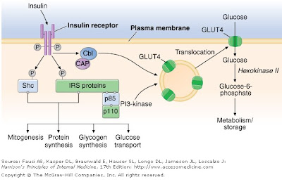
Once insulin is secreted into the portal venous system, ~50% is degraded by the liver. Unextracted insulin enters the systemic circulation where it binds to receptors in target sites. Insulin binding to its receptor stimulates intrinsic tyrosine kinase activity, leading to receptor autophosphorylation and the recruitment of intracellular signaling molecules, such as insulin receptor substrates (IRS) (Fig. 2). IRS and other adaptor proteins initiate a complex cascade of phosphorylation and dephosphorylation reactions, resulting in the widespread metabolic and mitogenic effects of insulin. As an example, activation of the phosphatidylinositol-3'-kinase (PI-3-kinase) pathway stimulates translocation of glucose transporters (e.g., GLUT4) to the cell surface, an event that is crucial for glucose uptake by skeletal muscle and fat. Activation of other insulin receptor signaling pathways induces glycogen synthesis, protein synthesis, lipogenesis, and regulation of various genes in insulin-responsive cells.
Figure 2. Insulin signal transduction pathway in skeletal muscle. The insulin receptor has intrinsic tyrosine kinase activity and interacts with insulin receptor substrates (IRS and Shc) proteins. A number of "docking" proteins bind to these cellular proteins and initiate the metabolic actions of insulin [GrB-2, SOS, SHP-2, p65, p110, and phosphatidylinositol-3'-kinase (PI-3-kinase)]. Insulin increases glucose transport through PI-3-kinase and the Cbl pathway, which promotes the translocation of intracellular vesicles containing GLUT4 glucose transporter to the plasma membrane. (Adapted from WL Lowe, in Principles of Molecular Medicine, JL Jameson (ed). Totowa, NJ, Humana, 1998; A Virkamaki et al: J Clin Invest 103:931, 1999. For additional details see AR Saltiel, CR Kahn: Nature 414:799, 2001.)
Glucose homeostasis reflects a balance between hepatic glucose production and peripheral glucose uptake and utilization. Insulin is the most important regulator of this metabolic equilibrium, but neural input, metabolic signals, and other hormones (e.g., glucagon) result in integrated control of glucose supply and utilization. In the fasting state, low insulin levels increase glucose production by promoting hepatic gluconeogenesis and glycogenolysis and reduce glucose uptake in insulin-sensitive tissues (skeletal muscle and fat), thereby promoting mobilization of stored precursors such as amino acids and free fatty acids (lipolysis). Glucagon, secreted by pancreatic alpha cells when blood glucose or insulin levels are low, stimulates glycogenolysis and gluconeogenesis by the liver and renal medulla. Postprandially, the glucose load elicits a rise in insulin and fall in glucagon, leading to a reversal of these processes. Insulin, an anabolic hormone, promotes the storage of carbohydrate and fat and protein synthesis. The major portion of postprandial glucose is utilized by skeletal muscle, an effect of insulin-stimulated glucose uptake. Other tissues, most notably the brain, utilize glucose in an insulin-independent fashion.


6 comments:
Good and simple for naive readers..! still u c'd add some good pics and expalin the exact Molecular mechanisms.
PJ
Thanks it was very useful for me
hi,I'm a medicine student,it was so useful for me,thx
ya its u little brief i think, since i am doing PG in medical biochemistry. Can u detail it some more sir
Hello, this weekend is good for me, since this time i am reading this enormous informative article here at my home. insulin like growth factor
I was diagnosed as HEPATITIS B carrier in 2013 with fibrosis of the
liver already present. I started on antiviral medications which
reduced the viral load initially. After a couple of years the virus
became resistant. I started on HEPATITIS B Herbal treatment from
ULTIMATE LIFE CLINIC (www.ultimatelifeclinic.com) in March, 2020. Their
treatment totally reversed the virus. I did another blood test after
the 6 months long treatment and tested negative to the virus. Amazing
treatment! This treatment is a breakthrough for all HBV carriers.
Post a Comment