Schizophrenia (Part 1)
S. Hossein Fatemi, MD, PhD, in The Medical Basis of Psychiatry, 3rd Ed. Humana Press. 2008.
1. History
Schizophrenia is a debilitating disease of the brain that has been described by various physicians for centuries . Hippocrates referred to paranoia as a potential antecedent of present day psychosis . Aretaeus of
However, the greatest and most methodological and comprehensive description of schizophrenia was heralded by the German psychiatrist, Emil Kraepelin, who referred to this disease as dementia praecox and separated it from manic depressive psychosis. Indeed, Kraepelin’s predecessor, Wilhelm Griesinger of
Eugen Bleuler, another leading figure in 20th century psychiatry, coined the term schizophrenia to describe the affective disturbance, the ambivalence and sense of isolation (autism), and associative (cognitive) disturbances observed in patients with dementia praecox. Bleuler also described schizophrenia as less of a dementing illness, with a more optimistic prognosis than Kraepelin had suggested. Finally, Kurt Schneider provided the concept of first- and second-rank symptoms (Table 1), to describe schizophrenia. It is now clear that, although these symptoms are helpful in diagnosis of schizophrenia, they are not specific to this disorder.
2. Current Diagnostic Criteria
The current criteria used presently to diagnose schizophrenia, is the product of years of empirical testing. The Diagnostic and Statistical Manual, 4th edition, text revision (DSM-IV-TR) criteria include:
A. Characteristic symptoms: Two (or more) of the following, each present for a significant portion of time during a 1-month period (or shorter if successfully treated):
1) delusions
2) hallucinations
3) disorganized speech (e.g., frequent derailment or incoherence)
4) grossly disorganized or catatonic behavior
5) negative symptoms, i.e., affective flattening, alogia, or avolition
Note: Only one Criterion A symptom is required if delusions are bizarre or hallucinations consist of a voice keeping up a running commentary on the person’s behavior or thoughts, or two or more voices conversing with each other.
Table 1. Schneider’s symptoms.
| First rank | Second rank |
| Audible thoughts | Depressiveoreuphoric mood changes |
| Voices heard arguing | Emotional blunting |
| Voices heard commenting on one’s actions | Perplexity |
| The experience of influences playing on the body | Sudden delusional ideas |
| Thought withdrawal and other interferences with thought | |
| Diffusion of thought | |
| Delusional perception | |
| Feelings, impulses, and volitional acts experienced as the work or influence of others | |
Schneider K. Clinical Psychopathology.
B. Social/occupational dysfunction: For a significant portion of the time since the onset of the disturbance, one or more major areas of functioning, such as work, interpersonal relations, or self-care are markedly below the level achieved before the onset (or when the onset is in childhood or adolescence, failure to achieve expected level of interpersonal, academic, or occupational achievement).
C. Duration: Continuous signs of the disturbance persist for at least 6 months. This 6-month period must include at least 1 month of symptoms (or shorter if successfully treated) that meet Criterion A (i.e., active-phase symptoms) and may include periods of prodromal or residual symptoms. During these prodromal or residual periods, the signs of the disturbance may be manifested by only negative symptoms or two or more symptoms listed in Criterion A present in an attenuated form (e.g., odd beliefs, unusual perceptual experiences).
D. Schizoaffective and mood disorder exclusion: Schizoaffec-tive disorder and mood disorder with psychotic features have been excluded because either 1) no major depressive, manic, or mixed episodes have occurred concurrently with the active-phase symptoms; or 2) if mood episodes have occurred during active-phase symptoms, their total duration has been brief relative to the duration of the active and residual periods.
E. Substance/general medical condition exclusion: The disturbance is not caused by the direct physiological effects of a substance (e.g., a drug of abuse, a medication) or a general medical condition.
F. Relationship to a pervasive developmental disorder: If there is a history of autistic disorder or another pervasive developmental disorder, the additional diagnosis of schizophrenia is made only if prominent delusions or hallucinations are also present for at least a month (or shorter if successfully treated).
3. Epidemiology
Schizophrenia affects 1% of the adult population in the world. The point prevalence of schizophrenia is approximately 5 per 1000 population and the incidence is approximately 0.2 per 1000 per year. This incidence rate was reported to be comparable in most societies; however, recent studies suggest greater variability. Schizophrenia has an earlier onset in male patients, with mean ages of onset of 20 and 25 years in male and female patients, respectively. Risk factors other than a familial history of schizophrenia include obstetric complications, parental age, prenatal infections, ethnicity, cannabis use, urbanicity, and modernization (trends toward a faster paced and more technological society).
4. Genetics
Emerging evidence points to schizophrenia as a familial disorder with a complex mode of inheritance and variable expression. Although single-gene disorders such as Huntington’s disease have homogenous etiologies, complex trait disorders such as schizophrenia have heterogeneous etiologies emanating from interactions between multiple genes and various environmental insults. Twin studies of schizophrenia suggest concordance rates of 45% for monozy-gotic twins and 14% for dizygotic twins. Consistent with this, a recent meta-analytic study showed a heritability of 81% for schizophrenia. Despite this high genetic predisposition, an 11% point estimate was suggested for the effects of environmental factors on liability to schizophrenia. Additionally, adoption studies show a lifetime prevalence of 9.4% in the adopted-away offspring of schizophrenic parents versus 1.2% in control adoptees. The adoption studies also clearly show that postnatal environmental factors do not play a major role in etiology of schizophrenia.
The mode of transmission in schizophrenia is unknown and most likely complex and non-Mendelian. Chromosomal abnormalities show evidence for involvement of a balanced reciprocal translocation between chromosomes 1q42 and 11q14.3, with disruption of disrupted in schizophrenia (DISC)-1 and DISC2 genes on 1q42 being associated with schizophrenia. Additionally, an association between a deletion on 22q11, schizophrenia, and velocardiofacial syndrome has been reported. Mice with similar deletions exhibit sensorimotor gating abnormalities.
Table 2. Risk genes for schizophrenia.
| Gene | Abbreviation | Locus |
| Neuregulin | NRG1 | 8p12-p21 |
| Dysbindin | DTNBP1 | 6p22 |
| G72 | G72 | 13q34 |
| d-amino acid oxidase | DAAO | 12q24 |
| RGS4 | RGS4 | 1q21-22 |
| Catechol-O-methyl | COMT | 22q11 |
| transferase | | |
| Proline dehydrogenase | PRODH | 22q11 |
| Reelin | RELN | 7q22 |
| Serotonin 2A receptor | 5HTR2A | 13q14-q21 |
From Sullivan et al., 2006; Fatemi et al., 2005; Wedenoja et al., 2007.
Linkage and association studies show 12 chromosomal regions containing 2,181 known genes and 9 specific genes being involved in the etiology of schizophrenia. Variations or polymorphisms in nine genes, including neuregulin 1 (NRG1), dystrobrevin-binding protein 1 (DTNBP1), G72 and G30, regulator of G protein signaling 4 (RGS4), catechol-O-methyltransferase (COMT), proline dehydrogenase (PRODH), DISC1 and DISC2, serotonin 2A receptor (HTR2A), and dopamine 3 receptor (DRD3) have been associated with schizophrenia (Table 2).
Table 3. Candidate genes: postmortem studies and animal models.
| Gene | Abbreviation | Postmortem | Animal model |
| Adenosine A2A receptor | ADORA2A | + | + |
| Apolipoprotein D | APOD | + | + |
| CDC42 guanine nucleotide exchange factor 9 | ARHGEF9 | + | |
| Complexin 2 | CPLX2 | + | + |
| Distal-less homeobox 1 | DLX1 | + | |
| Dopamine receptor D1 | DRD1 | + | |
| Dopamine receptor D2 | DRD2 | + | + |
| GABAA receptor, subunit A1 | GABRal | + | |
| GABAA receptor, subunit A5 | GABRa5 | + | + |
| GABAB receptor 1 | GABBR1 | + | |
| Glutamic acid decarboxylase 2 | GAD2 | + | |
| Glial fibrillary acidic protein | G FA P | + | + |
| Glutamate receptor, ionotropic, AMPA1 | GRIA1 | + | |
| Glutamate receptor, ionotropic, AMPA2 | GRIA2 | + | |
| Myelin and lymphocyte protein | MAL | + | |
| Myelin basic protein | MBP | + | + |
| Neuronal PAS domain protein 1 | NPAS1 | + | + |
| Proteolipid protein | PLP1 | + | |
| Reelin | RELN | + | + |
| Regulator of G protein signaling 4 | RGS4 | + | |
| Short stature homeobox 2 | SHOX2 | + | |
| Synapsin 2 | SYN2 | + | |
From Le-Niculescu et al., 2007 (14).
Anothe means of studying the genetic basis of schizophrenia uses the technique of DNA microarray. These studies are based on discovering genes either repressed or stimulated significantly in well-characterized postmortem brain tissues from subjects with schizophrenia and matched healthy control subjects; peripheral lymphocytes obtained from schizophrenic and matched healthy controls and antipsychotic-treated brains of rodents (Table 3). Genes involved in drug response, or in etiopathogenesis of schizophrenia can be compared and studied to better understand the mechanisms responsible for this illness.
5. Etiology
The concept of schizophrenia as a neurodevelopmental disease dates back to the period of Kraepelin and Bleuler. Early manifestations of disease as exemplified by premorbid signs and deficits in social interaction were observed by Kraepelin and Bleuler in children who later developed schizophrenia. Later, Southard reported on the presence of neuropathological signs in brains of subjects with schizophrenia that further pointed to maldevelopmental origins of this disorder.
5.1. Neurochemistry of Schizophrenia
5.1.1. The Dopamine Hypothesis
Dopaminergic tracts are composed of four branches: 1) the nigrostriatal tract, originating from the substantia nigra and ending in the dorsal striatum, deals with initiation of movement, motor control, sensorimotor coordination, cognitive integration, and habituation; 2) the mesolimbic tract, originating from the ventral tegmental area and ending in hippocampus, amygdala, and ventral striatum, deals with cognitive/attentional, motivational, and reward systems; 3) the mesocortical tract, originating from the ventral tegmental area and ending in the cortical structures, deals with attention, motivation, and reward systems; and 4) the tuberoin-fundibular tract, the cell bodies originating from the arcuate nucleus and periventricular hypothalamic areas and ending in the infundibulum and anterior pituitary, dealing with control of prolactin release. The dopamine (DA) receptors are classified into two distinct families of D1-like (D1 and D5) and D2-like (D2, D3, D4) receptors (25). The D1 receptors are localized to prefrontal cortex (PFC) and striatum. The D2 receptors are localized mostly to striatum, but with lower concentrations in the hippocampus, amygdala, and entorhinal cortex. The D3 receptors are localized to the ventral striatum. The D4 receptors are present in the hippocampus and PFC. Finally, the D5 receptors are found in the hippocampus and entorhinal cortex. Presynaptic dopamine receptors such as D2 and D3 are localized to cell bodies or axon terminals of neurons. Dopamine helps in modulating glutamatergic inputs and pyramidal cell excitability.
The dopamine hypothesis of schizophrenia is based on the assumption that dopamine hyperactivity causes psychotic symptoms, and that dopamine antagonists such as chlorpromazine treat the psychotic symptoms. Additionally, administration of d-amphetamine to healthy volunteers leads to production of psychotic symptoms and worsens psychosis in schizophrenic subjects. One limitation of this hypothesis is that hallucinogens such as LSD or psilocybin (acting on the serotonin system) or dissociative anesthetics such as ketamine or phencyclidine (PCP) (acting on the glutamate system) also cause psychotic symptoms. A further limitation of this hypothesis is that consistent abnormalities have not been found in dopamine receptors or dopamine metabolites in subjects with schizophrenia. The two consistent postmortem findings include an increase in D2-like receptors in striatum of schizophrenic patients and lack of changes in striatal densities of D1 receptors or dopamine transporters. However, a recent finding of upregulated D1 receptor binding in the dorsolateral PFC (DLPFC) of schizophrenic subjects has been associated with impaired working memory performance.
5.1.2. The Serotonin Hypothesis
The serotonin (5HT) neurons emanate from the midbrain dorsal and median raphe nuclei and project to several sites including hippocampus, striatum, and cortex. The number of various serotonin receptor types in the brain exceed 15, with the most important receptors being 5HT1, 5HT2, 5HT3, 5HT6, and 5HT7. Inhibitory somatodendritic serotonin autoreceptors (5HT1A) are localized to raphe seroton-ergic neurons, which, on activation, lead to decreased firing of the neurons. In contrast, terminal autoreceptors modulate synthesis and release of serotonin. 5HT3 receptors help to stimulate dopamine release. Additionally, pyramidal cells in the mesocortical areas bear postsynaptic 5HT2A receptors that subserve serotonin–dopamine interaction in various brain areas.
Although LSD and other serotonergic agonists can lead to psychotic symptoms in healthy individuals, the latter symptoms consist mostly of visual hallucinations, which are less frequently seen in schizophrenic patients. Despite the shortcomings of the serotonin hypothesis of schizophrenia, the atypical antipsychotic agents used extensively today are potent antagonists of the 5HT2 receptors, which may help in treating negative symptoms of schizophrenia and reduce extrapyramidal side effects (EPS).
5.1.3. The Glutamate Hypothesis
Glutamate is the main excitatory neurotransmitter in the central nervous system (CNS). Approximately 60% of neurons and 40% of synapses of the brain are glutamatergic in nature. The glutamate receptors consist of ionotropic and metabotropic families. The ionotropic glutamate receptors (those working through Ca++ channels) include N-methyl-d-aspartic acid (NMDA), α-amino-3-hydroxy-5-methyl-4-isoxazolepropionic (AMPA), and kainate receptors. The metabotropic family (receptors that indirectly regulate electrical signaling by activation of various second messengers) consists of groups I, II, and III receptors. The glutamatergic hypothesis of schizophrenia is based on deceased levels of glutamate in the cerebrospinal fluid (CSF) of schizophrenic subjects and decreased expression of NMDA and AMPA receptors in the hippocampus and thalamus of schizophrenic subjects. Additionally, use of noncompetitive and competitive antagonists of NMDA receptors can lead to production of positive, negative, and cognitive symptoms of schizophrenia. Administration of clozapine can block the NMDA antagonistic effects of PCP. Several compounds, such as glycine, d-serine, and d-cycloserine, have been reported to reduce positive and negative symptoms in subjects with schizophrenia. Genetic evidence also points to involvement of several genes (DAAO, G72, neuregulin, dysbindin, RGS4) that impact the glutamate system in schizophrenia. Mice with alterations in NMDA receptors show hyperactivity and schizophrenic-like behaviors.
5.1.4. The GABAergic Hypothesis
Gamma-aminobutyric acid (GABA) is the major inhibitory neurotransmitter in the mammalian brain. Recent postmortem evidence suggests involvement of glutamic acid decarboxy-lase 65- and 67-kDa proteins (GAD65 and 67), the rate-limiting enzymes that convert glutamate to GABA, in the cerebellum of schizophrenic subjects. Supportive data also point to decreases in GAD67 species in brains of subjects with schizophrenia. Furthermore, Reelin, an important factor involved in synaptic plasticity that colocalizes to GABAergic interneurons is reduced in brains of subjects with schizophrenia (42, 46–48)
5.2. Neurodevelopmental Theories of Schizophrenia
The accumulation of a large body of evidence during the last century points to involvement of pathologic processes that begin in utero and lead to development of schizophrenia in adolescence. These neurodevelopmental abnormalities, beginning in utero, as early as late first or early second trimester, have been suggested to lead to activation of pathologic neural circuits during adolescence, which may underlie development of psychotic symptoms in the susceptible individual.
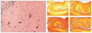 Figure 1. a Several Reelin-positive cells are localized to the hilus (CA4) of hippocampal complex. M, dentate inner molecular layer, GC, granular cell layer. Original magnification, ×40. b SNAP-25 immunostaining is localized to various layers of ventral hippocampus in subjects with bipolar disorder (A), major depression (B), and schizophrenia (D) compared with a normal control subject (C). Note the diminution in SNAP-25-specific immunostaining in the strata oriens and moleculare of patients with bipolar disorder (A) and schizophrenia (D) versus an increase in SNAP-25 depressed (B) and normal levels in control (C) subjects. The following hippocampal fields were identified and analyzed by densitometry: from the outside in, alveolus, stratum oriens, strata pyramidale, radiatum and lucidum combined, stratum moleculare (combined hippocampal layer as well as outer and inner dentate molecular layers), stratum granulosum, cornu ammonis (CA4) or hilus, cornu ammonis 3 (CA3), cornu ammonis 2 (CA2), subiculum, and presubiculum. (a Reprinted with permission from the Nature Publishing Group (47). b Reprinted with permission from Lippincott Williams and Wilkins)
5.2.1. Obstetric and Perinatal Complications
There is a large body of epidemiologic research showing an increased frequency of obstetric and perinatal complications in schizophrenic patients. The complications observed include periventricular hemorrhages, hypoxia, and ischemic injuries.
5.2.2. Brain Structural Studies
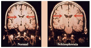
A consistent observation in schizophrenia is the enlargement of the cerebroventricular system. The abnormalities are present at onset of disease, progress very slowly, and are unrelated to the duration of illness or treatment regimen. Additionally, cerebroventricular enlargement distinguishes affected from unaffected discordant monozygotic twins (Fig. 2). Furthermore, gross brain abnormalities have been identified in the DLPFC, hippocampus, cingulate cortex, and superior temporal gyrus. Some reports also indicate presence of brain structural abnormalities in individuals at high risk for development of schizophrenia and in unaffected first-degree relatives of subjects with schizophrenia. More recently, studies of white matter tracts show evidence of disorganization and lack of alignment in white fiber bundles in frontal and temporoparietal brain regions in schizophrenia (Fig. 2).
5.2.3. Postmortem Histologic Studies
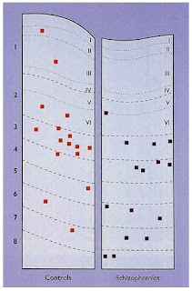
Numerous reports have documented the presence of various neuropathologic findings in postmortem brains of patients with schizophrenia. These findings consist of cortical atrophy; ventricular enlargement; reduced volumes of hippocampus, amygdala and parahippocampal gyrus; disturbed cytoarchitecture in the hippocampus; cell loss and volume reduction in the thalamus; abnormal translocation of NADPH–diaphorase-positive cells in frontal and hippocampal areas (Fig. 3); and reduced cell size in Purkinje cells of the cerebellum.
However, by far the greatest abnormalities have been found in the prefrontal, ventral hippocampal, and cerebellar cortices of schizophrenic brains (61). Collectively, these data reflect abnormal corticogenesis during the mid-gestation period in schizophrenic patients. Additionally, several recent reports using magnetic resonance imaging (MRI) and diffusion tensor imaging (DTI) techniques have shown reduced white matter diffusion anisotropy (diffusion changes in water in white matter) in subjects with schizophrenia. In brain white matter, water diffusion is highly anisotropic, with greater diffusion in the direction parallel to the axonal tracts. Thus, reduced anisotropy of water diffusion has been proposed to reflect compromised white matter integrity in schizophrenia. Furthermore, reductions in white matter anisotropy reflect disrupted white matter connections, which supports the disconnection model of schizophrenia. Reduced white matter diffusion anisotrophy has been observed in prefrontal, parieto-occipital, splenium of corpus callosum, arcuate and uncinate fasciculus, parahippocampal gyri, and deep frontal perigenual regions of brain in schizophrenic patients. There are also negative findings showing no white matter abnormalities in schizophrenia. It is conceivable that downregulation of genes affecting production of myelin-related proteins as well as other components of axons may lay the foundation for white matter abnormalities that develop later in life in subjects who become schizophrenic. Several recent reports now indicate that either glial cells are dysfunctional or unaffected in schizophrenia. Thus, absence of gliosis in brains of schizophrenic subjects may no longer imply direct support for initiation of early insults in utero in these patients.
Figure 2. Ventricular size in monozygotic twins discordant for schizophrenia. Coronal MRI scans of twins discordant for schizophrenia show lateral ventricular enlargement in the affected twin (reprinted with permission from the Massachusetts Medical Society (179). All rights reserved).
Figure 3. These camera lucida drawings compare the distribution of nicotinamide–adenine dinucleotide phosphate–diaphorase-stained neurons (squares) in sections through the superior frontal gyrus of a control and schizophrenic brain. There is a significant shift in the direction of the diaphorase positive neurons in the white matter in the schizophrenic brain. Numbers 1 to 8 indicate compartments of the brain; Roman numerals indicate the cortical layers (reprinted with permission from the American Medical Association (60). All rights reserved)
5.2.4. Biochemical Brain Marker Anomalies and DNA Microarray Studies of Schizophrenia
Biological markers consistent with prenatal occurrence of neurodevelopmental insults in schizophrenia include changes in the normal expression of proteins that are involved in early migration of neurons and glia, cell proliferation, axonal outgrowth, synaptogenesis, and apoptosis (Table 4). Some of these markers have been investigated in studies of various prenatal insults in potential animal models for schizophrenia, thus, helping in deciphering the molecular mechanisms for genesis of schizophrenia (Table 3).
Several recent reports implicate various gene families as being involved in pathology of schizophrenia using DNA microarray technology, i.e., genes involved in signal transduc-tion, cell growth and migration, myelination, regulation of presynaptic membrane function, and GABAergic function. By far the most well studied and replicated data deal with genes involved in oligodendro-cyte and myelin-related functions. Hakak et al., using mostly elderly schizophrenic and matched control DLPFC homogenates, showed downregulation of five genes whose expression is enriched in myelin-forming oligodendrocytes, which have been implicated in the formation and maintenance of myelin sheaths. Later, Tkachev et al., using area 9 homogenates from the Stanley Brain collection showed significant downregulation in several myelin and oligoden-drocyte related genes, such as proteolipid protein 1, myelin associated glycoprotein (MAG), oligodendrocyte-specific protein CLDN11, myelin oligodendrocyte glyco-protein (MOG), myelin basic protein (MBP), neuroregulin receptor ERBB3, transferrin, olig 1, olig 2, and Sry Box10 (SOX-10). Mirnics et al. showed downregulation of genes involved in presynaptic function in the PFC, such as methyl maleimide-sensitive factor, synapsin II, synaptojanin 1, and synaptotagmin 5. Vawter et al. showed down-regulation of histidine triad nucleotide-binding protein and ubiquitin-conjugating enzyme E2N. Another important family of genes involved in schizophrenia is genes involved in gluta-mate and GABAergic function. Hakak et al. showed an upregulation of several genes involved in GABA transmission, such as GAD65 and GAD67. However, several reports have shown decreases in these proteins in schizophrenia. Hashimoto et al. showed a downregulation of Parval-bumin gene, and Vawter et al. showed downregulation of glutamate receptor AMPA 2. Another gene family of import in schizophrenia deals with signal transduction. Hakak et al. showed upregulation of several postsynaptic signal transduc-tion pathways known to be regulated by dopamine, consistent with the dopamine hypothesis of schizophrenia, such as protein kinase A R II and NT-related protein 2. In a similar vein, Mirnics et al. also showed downregulation of the RGS4 gene in the PFC of patients with schizophrenia. Finally, Chung et al. showed upregulation of the heat shock 70 gene in schizophrenic brain.
5.2.5. Effects of Adverse Environmental Events on Brain Development In Utero
There is ample evidence to indicate that the greatest risk factor for development of schizophrenia is being related to a person with schizophrenia, i.e., in some subgroups, heredity can explain more than 80% of the liability to schizophrenia. However, there is also a robust collection of reports indicating that environmental factors, especially viral infections, can increase the risk for development of schizophrenia. Emil Kraepelin referred to potential for infections causing some forms of dementia praecox (schizophrenia) during early stages of brain development. Meninger described 67 cases of schizophrenia in a large cohort of patients who contracted influenza during the pandemic of 1919. Later, Hare et al. and Machon et al. reported on an excess of schizophrenic patients being born during late winter and spring as indicators of potential influenza infections being responsible for these cases. Indeed, the majority of nearly 50 studies performed in the intervening years indicate that a 5 to 15% excess of schizophrenic births in the
Northern Hemisphere occurs during the months of January and March. This excess winter birth has not been shown to be caused by unusual patterns of conception in mothers or to a methodological artifact. Machon et al. and Mednick et al. showed that the risk of schizophrenia was increased by 50% in Finnish individuals whose mothers had been exposed to the 1957 A2 influenza during the second trimester of pregnancy. Later, 9 of 15 studies performed replicated Mednick’s findings of a positive association between prenatal influenza exposure and schizophrenia. These association studies showed that exposure during the 4th to 7th months of gestation affords a window of opportunity for influenza virus to cause its terato-genic effects on the embryonic brain. Additionally, three out of five cohort and case–control studies support a positive association between schizophrenia and maternal exposure to influenza prenatally. Subsequent studies have now shown that other viruses, such as rubella, may also increase the risk for development of schizophrenia in the affected progeny of exposed mothers. By far, the most exciting evidence linking viral exposure to development of schizophrenia was published recently by Karlsson et al., who provided data suggestive of a possible role for retroviruses in the pathogenesis of schizophrenia. Karlsson and colleagues identified nucleotide sequences homologous to retroviral polymerase genes in the CSF of 28.6% of subjects with schizophrenia of recent origin and in 5% of subjects with chronic schizophrenia. In contrast, such retroviral sequences were not found in any individuals with noninflammatory neurological illnesses or in healthy subjects. The upshot of these studies and previous epidemiological reports is that schizophrenia may represent the shared phenotype of a group of disorders whose etiopathogenesis involves the interaction between genetic influences and environmental risks, such as viruses operating on brain maturational processes. Moreover, identification of potential environmental risk factors, such as influenza virus, or retroviruses such as endogenous retroviral-9 family and the human endogenous retrovirus-W species observed by Karlsson et al., will help in targeting early interventions at repressing the expression of these transcripts. An alternate approach would be to vaccinate against influenza, thus, influencing the course and outcome of schizophrenia in the susceptible individuals.
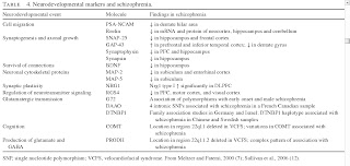
Table 4. Neurodevelopmental markers and schizophrenia (click image to view)
At least two mechanisms may be responsible for transmission of viral effects from the mother to the fetus: 1) Via direct viral infection. There are clinical, as well as direct experimental reports showing that human influenza A viral infection of a pregnant mother may cause transplacental passage of viral load to the fetus. In a series of reports, Aronsson and colleagues used human influenza virus (A/WSN/33, a neurotropic strain of influenza A virus), on day 14 of pregnancy, to infect pregnant C57BL/6 mice intranasally. Viral RNA and nucleoprotein were detected in fetal brains and viral RNA persisted in the brains of exposed offspring for at least 90 days of postnatal life, thus, showing evidence for transplacental passage of influenza virus in mice and the persistence of viral components in the brains of progeny into young adulthood. Additionally, Aronsson et al. demonstrated that 10 to 17 months after injection of the human influenza A virus into olfactory bulbs of TAP1 mutant mice, viral RNA encoding the nonstructural NS1 protein was detected in midbrain of the exposed mice. The product of the NS1 gene is known to play a regulatory role in the host–cell metabolism. Several in vitro studies have also shown the ability of human influenza A virus to infect Schwann cells, astrocytes, microglial cells, and neurons, and hippocampal GABAergic cells, selectively causing persistent infection of target cells in the brain. 2) Via induction of cytokine production. Multiple clinical and experimental reports show the ability of human influenza infection to induce production of systemic cytokines by the maternal immune system, the placenta, or even the fetus itself. New reports show the presence of serologic evidence of maternal exposure to influenza as causing increased risk of schizophrenia in offspring. Offspring of mothers with elevated IgG and IgM levels, as well as antibodies to herpes simplex virus type 2 during pregnancy, have an increased risk for schizophrenia. Cytokines such as inter-leukin (IL)-1(3, IL-6, and tumor necrosis factor (TNF)-a are elevated in pregnant mothers after maternal infection and after infection in animal models. All of these cytokines are known to regulate normal brain development and have been implicated in abnormal corticogenesis. Additionally, expression of messenger RNAs (mRNAs) for cytokines in the CNS is developmentally regulated both in man and in mouse, emphasizing the significant role that cytokines play during neurodevelopment. IL-1(3, IL-6 and TNFα cross the placenta and are synthesized by mother, by the placenta, and by the fetus. Maternal levels of TNFα and IL-8 have been shown to be elevated in human pregnancies in which the offspring goes on to develop schizophrenia. A more relevant series of studies in different animal models for schizophrenia show that maternal infection with human influenza mimic poly I:C, a synthetic double-stranded RNA that stimulates a cytokine response in mice, can cause abnormalities in prepulse inhibition, or, after maternal exposure to Escherichia coli cell wall endotoxin lipopolysaccharide, cause disruption of sensorimotor gating in the offspring. Finally, maternal exposure to poly I:C also causes disrupted latent inhibition in rats. All of these models suggest that direct stimulation of cytokine production by infections or immunogenic agents cause disruptions in various brain structural or behavioral indices of relevance to schizophrenia. Other factors associated with increased schizophrenic births include famine during pregnancy, Rh factor incompatibility, and autoimmunity caused by infectious agents.
Table 5. Neurologic soft signs.
Choreoathetoid movements in preschizophrenic children
Abnormal gait
Abnormal body movements
Mannerisms
Grimacing
Stereotypies
Abnormal reflexes
Increase/decrease muscle tone
Abnormal rapid eye movements (saccades)
Frequent blinking
Dysdiadochokinesia
Astereognosis
Poor right-left discrimination
Anosognosia
Apraxia
Sympathetic arousal
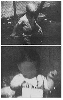
Figure 4. Abnormalities of left-hand posture in preschizophrenic children (reprinted with permission from the Schizophrenia Bulletin ).
5.2.6. Congenital Anomalies and Developmental Dysfunction
Multiple markers of congenital anomalies indicative of neurodevelopmental insults have been found in schizophrenia. Such anomalies include agenesis of corpus callosum, stenosis of sylvian aqueduct, cerebral hamartomas, and cavum septum pellucidum. Presence of low-set ears, epicanthal eye folds, and wide spaces between the first and second toes, are suggestive of first trimester anomalies. There is, however, support for abnormal dermatoglyphics in patients with schizophrenia, indicating a second trimester event. Multiple reports indicate the presence of premorbid neurologic soft signs (Table 5) in children who later develop schizophrenia. Slight posturing of hands and transient choreoathetoid movements has been observed during the first 2 years of life in children who later developed schizophrenia (Fig. 4). Additionally, poor performance on tests of attention and neuromotor performance, mood and social impairment, and excessive anxiety have been reported to occur more frequently in high-risk children with a schizophrenic parent. All of these findings are consistent with schizophrenia as a syndrome of abnormal brain development.
6. Clinical Findings
6.1. Clinical Signs and Symptoms of Schizophrenia
The current diagnostic criteria adopted by DSM-IV-TR is based on extensive research dating back to initial findings of Kraepelin, Bleuler, and Schneider. Unlike other medical conditions, no one sign is diagnostic of schizophrenia. Thus, it is absolutely important to obtain as much clinical history regarding the patient to help establish a correct diagnosis.
As mentioned earlier, The DSM-IV-TR criteria consist of six major topics:
1. Criterion A includes two or more of the following symptoms during a 1-month period: 1) delusions, 2) hallucinations, 3) disorganized speech, 4) grossly disorganized or catatonic behavior, 5) negative symptoms (flat affect, alogia, or avolition). Alternatively, the diagnosis of schizophrenia may be based on presence of bizarre delusions alone, auditory hallucinations of a voice keeping a running commentary one one’s daily activities, or two or more voices conversing with each other. Delusions are fixed false beliefs not congruent with one’s cultural or religious background. Schizophrenic patients may exhibit delusions that correspond to themes of persecution, grandiosity, outside control, guilt, thought broadcasting, thought withdrawal/insertion, or ideas of reference. Bizarre delusions are highly implausible false beliefs. Hallucinations are abnormal perceptions of sensory experiences that occur in the absence of external stimuli. Hallucinations can be based on various types of sensory modalities, such as auditory, visual, gustatory, olfactory, tactile, or cenesthetic (change in the normal quality of feeling tone in a part of the body). Auditory hallucinations are more common in schizophrenic subjects, and occurrence of other types of hallucinations should be considered as potential signs of other medical/organic etiologies. Command auditory hallucinations may lead the patient to act on the command to harm self or others. Disorganized speech and behavior reflect underlying thought disorder or impairment. Examples of abnormal speech include circumstantiality, tangentiality, derailment, illogicality, incoherence, concrete speech, clanging, neologisms, echolalia, thought blocking, perseverations, and poverty of content. Disorganized behavior includes bizarre postures, stereotyped behavior, echopraxia, negativism, catatonic stupor/excitation, waxy flexibility, unprovoked outbursts of laughter or violent behavior, severe neglect of hygiene, poor self-care and grooming, grimacing, athetosis, and mutism. Grossly disorganized or catatonic behavior may also include verbigeration, primitive reflexes, autonomic hyperactivity, staring, and rigidity. Finally, negative symptoms reflect deficits of normal functions and examples include affective flattening, avolition, alogia, anhedonia, social withdrawal, and diminished capacity to feel close to others. These negative symptoms reflect endogenous markers of schizophrenia and are, thus, called primary negative symptoms. Negative symptoms such as depression or demoralization, which may be caused by side effects of medications, are called secondary negative symptoms.
2. Criterion B reflects significant social/occupational dysfunction, because the onset of disease is such that patient is not able to work, go to school, provide self-care, and relate to others normally.
3. Criterion C reflects the continuous presence of illness for at least 6 months. This 6-month period must include 1 month of Criterion A (active-phase symptoms), and may include prodromal or residual symptoms. The prodromal symptoms predate the active phase of psychosis and consist of symptoms of impending social withdrawal, restricted range of affect, cognitive difficulties, and increasing emergence of odd behavior. The residual phase of the disease reflects presence of attenuated forms of positive or negative symptoms.
4. Criterion D. Presence of mood disorders or schizoaffective disorder has been excluded.
5. Criterion E. The schizophrenic symptoms are not caused by various medical conditions or by direct physiological effects of a drug of abuse.
6. Criterion F. In the presence of autism or another pervasive developmental disorder, a diagnosis of schizophrenia is made based on the presence of prominent delusions or hallucinations for at least 1 month.
6.2. Mental Status Examination in a Subject with Schizophrenia
6.2.1. Appearance
On examination, a patient with possible diagnosis of schizophrenia may appear disheveled, and exhibit evidence of poor self-care or grooming. The patient may appear suspicious, relate poorly to the examiner, and exhibit bizarre postures, stereotypy, grimacing, athetosis, mutism, or catatonic agitation or stupor. The clinician must look for presence of abnormal EPS, such as dystonia, tardive dyskinesia, rabbit syndrome, or akathisia.
6.2.2. Affect
The subject may exhibit an affect that is incongruent with patient’s state of mind or is described as inappropriate. Affect may also appear blunted, constricted, or flat. However, absence of a sad affect does not exclude presence of a depressed mood. Flat affect may be secondary to drug-induced parkinsonism or EPS.
6.2.3. Mood
Mood may be depressed or variable. Because depression occurs frequently in schizophrenic subjects and causes a high rate of suicide, it is essential for clinicians to evaluate for the presence of depression and to treat it.
6.2.4. Speech
Evaluation of patient’s speech may identify presence of loose, illogical, or bizarre thought patterns. Additionally, patients may exhibit various abnormalities of speech, such as tangentiality, circumstantiality, neologisms, clang association (speech directed by the sound of a word rather than by its meaning), perseveration, and poverty of content. Patients may also express echolalia or thought blocking. Evidence of tardive dyskinesia or dystonia affecting a patient’s speech should be investigated.
6.2.5. Thought Form and Content
Thought form can be ascertained while listening to patient’s speech. Thus, presence of a loose and illogical thought pattern is tantamount to evidence of formal thought disorder. Also, answering questions inappropriately (for example tangential responses: Q: What color is the sky? A: It rained yesterday) indicates a formal thought disorder. Alternatively, thought content may be replete with evidence for delusions, ideas of reference, thought broadcasting, thought insertion or withdrawal, ideas of persecution or grandiosity, or outside control. It is imperative to evaluate the patient for the presence of depression, mania, anxiety, panic, racing thoughts, irritability, suicidal and homicidal ideations, or plans or histories of suicide or violence toward others. Presence of obsessive– compulsive symptoms, past or recent histories of traumatic events, and signs of posttraumatic stress disorder (PTSD) should be evaluated.
6.2.6. Perceptual Abnormalities
Presence of hallucinations, illusions, de´ja vu, depersonalization, and derealization should be determined. Intensity, frequency and past or present occurrence of various types of hallucinations should be determined. For example, the specifics of the sex of voices heard, the loudness, and the origin of voices emanating from inside or outside the patient’s head should be investigated. The presence of command auditory hallucinations that may order patients to harm themselves or others should be ascertained. The form and color of visual hallucinations should be evaluated. Visual hallucinations occurring during sleep, before, or immediately after sleep are not necessarily indicative of a pathological process.
6.2.7. Cognition/Sensorium
Subjects with schizophrenia generally present with an intact sensorium, i.e., they are alert and oriented to place, person, and time. Evaluation of immediate, short-, and long-term memory and attention should be performed. Other cognitive domains may present major abnormalities, either through bedside examination or by performance of a neuropsycho-logical battery of tests. Insight and judgement are generally evaluated by questioning a patient’s awareness of their illness or their ability to interact normally with others, respectively. These mental abilities are more likely to be impaired in a patient with schizophrenia. Use of proverb analysis and similarities may shed light on a patient’s degree of abstraction or concreteness of thought. Most schizophrenic patients exhibit IQ scores 10 points lower than the general public and exhibit impairments in attention, working memory, visual spatial memory, semantic memory, recall memory, and executive functions. The presence of cognitive abnormalities in schizophrenia seems to be independent of positive, negative, or disorganization symptoms.
6.2.8. Physical Examination
As in all other fields of medicine, every psychiatrist must be able to perform a focused physical examination to identify the presence of any general medical or neurologic abnormalities. However, because of potential boundary issues, the presence of a nurse or a chaperone in the examination room is warranted. Patients with schizophrenia have a higher burden of medical comorbidities and, thus, should be evaluated fully. Review of systems should cover neurologic, cardiovascular, ear, nose, and throat, gastrointestinal, genitourinary, derma-tologic, and endocrine systems. The presence of neurological soft signs (Table 5), such as abnormal gait, abnormal reflexes, changed muscle tone, abnormal rapid eye movements (saccades), frequent blinking, dysdiadochokinesia, astereog-nosis, poor right–left discrimination, anosognosia, apraxia, sympathetic arousal, choreoathetosis, mannerisms, grimacing, stereotypies, and abnormal body movements are indicators of the neurodevelopmental origins of schizophrenia. Because of increased risk for diabetes and metabolic syndrome caused by atypical agents, it would be judicious to obtain baseline and follow-up weight, vital signs, and waist circumference (body mass index) for every patient examined.
6.2.9. Neuropsychological Testing
Formal objective tests, such as the Halstead–Reitan Battery, the Luria–Nebraska Battery, the Wechsler Adult Intelligence Scale, the Wechsler Intelligence Scale for Children, the Wisconsin Card Sorting Test (WCST), the Brief Assessment of Cognition in Schizophrenia (BACS), and the Measurement and Treatment Research to Improve Cognition in Schizophrenia (MATRICS) can identify various brain abnormalities and should be performed in all initial evaluations.
6.2.10. Laboratory Investigations
6.2.10.1. Blood, Urine, and CSF
Although no specific blood, urine, or CSF tests are available to demonstrate the presence of schizophrenia unequivocally, performance of certain blood and urine tests to exclude the presence of nonpsychiatric causes for psychotic symptoms is warranted in all initial examinations. Tests such as thyroid function (thyroid-stimulating hormone [TSH], T3, T4), rapid plasma reagin [RPR], HIV, fasting lipid panel, complete blood count with differential, metabolic panel (electrolytes, liver enzymes, blood urea nitrogen [BUN], glucose, and creati-nine), erythrocyte sedimentation rate (ESR), antinuclear antibody (ANA), rheumatoid factor (RF), B12, folate, prolactin, urinalysis, urine tox screen, blood alcohol level, and baseline electrocardiogram (ECG) should be performed during the initial evaluation, as warranted by clinical judgment of the examining psychiatrists. Evaluation of hemoglobin A1C to exclude the presence of diabetes mellitus at baseline and after institution of atypical antipsychotics is warranted. In certain cases, when suspicion of infectious etiologies is present, referral of patient to a neurologist for a lumbar puncture is warranted.
6.2.10.2. Brain Imaging
Ventricular enlargement and increased brain sulcal prominence can be identified in computed tomography (CT) or MRI views. Although these findings are supportive of brain abnormality, they are not specific to schizophrenia. Additionally, despite identification of a number of other brain abnormalities, such as smaller cerebral and cranial size, smaller medial temporal structures (hippocampus), enlargement of lenticular nucleus, cerebellar vermal dysplasia, and smaller thalamus in subjects with schizophrenia, none are diagnostic of schizophrenia. More recently, DTI has shown the presence of white matter tract abnormalities in schizophrenic patients, but, again, these findings have yet to be fully replicated.
6.2.10.3. Functional Brain Imaging
Use of functional MRI (fMRI), single-photon emission computed tomography (SPECT), and positron emission tomography (PET) scanning has provided a wealth of information regarding various brain function abnormalities in schizophrenia. These abnormalities include dysfunction of information storage and retrieval by the DLPFC, abnormal inhibitory response to sensory stimuli by the anterior cingulate cortex, abnormal encoding and retrieval of memory by the hippocampus, abnormal reception and integration of sensory information by the thalamic nuclei, primary sensory cortices, and the multimodal cortices, and impaired performance of cognitive tasks by the basal ganglia, thalamus, and cerebellum. Very recently, Hubl et al. showed that abnormal activation of the primary auditory cortex in schizophrenic subjects may be a biological cause for the production of auditory hallucination.
6.3. Differential Diagnosis
Psychotic symptoms are present in a number of conditions and must be excluded before entertaining the diagnosis of schizophrenia.
6.3.1. Mood Disorders
Psychotic symptoms, such as mood-congruent hallucinations or delusions may occur in severe depression or mania. Acquisition of a detailed history and correlative clinical data should guide the clinician regarding whether these symptoms occur because of mood abnormalities. Exclusive co-occurrence of psychotic symptoms with mood symptoms should denote the presence of a mood disorder with psychotic features. Alternatively, an uninterrupted period of illness, during which, at some time, there is a major depressive episode, a manic episode, or a mixed episode concurrent with symptoms that meet Criterion A for schizophrenia should point to the possibility of schizoaffective disorder.
6.3.2. Psychotic Disorders Caused by Medical, Neurologic, or Substance-Induced Conditions
The chronology of psychotic symptoms occurring in relation to an inciting condition or in association with physical or laboratory signs indicative of a medical or neurologic disorder is helpful in distinguishing nonschizophrenic psychosis. Many substances can cause psychosis, such as amphetamines, substituted amphetamines (ecstacy), hallucinogens, alcohol, barbiturates, cocaine, ketamine, PCP, and belladonna alkaloids. Examples of medical or neurologic conditions that induce psychosis include infectious causes (herpes encephalitis, neurosyphilis, AIDS), metabolic events (acute intermittent porphyria, vitamin B12 deficiency, carbon monoxide poisoning, homocystin-uria, heavy metal poisoning), neurologic events (temporal lobe epilepsy, frontal or limbic trauma, cerebrovascular accidents, Huntington’s disease, metachromatic leukodys-trophy, normal-pressure hydrocephalus, Wernicke–Korsakoff syndrome, Wilson’s disease, Creutzfeldt–Jakob disease), and various conditions, such as neoplasms, Fabry’s disease, Fahr’s disease, Hallervorden–Spatz disease, and systemic lupus erythematosus. Clearly, obtaining historical details on clinical course, performing physical examination, and doing pertinent laboratory examinations will help the psychiatrist in identifying the cause of psychotic symptoms and excluding schizophrenia.
6.3.3. Other Psychotic Disorders
Psychotic symptoms may occur during a period of mood abnormality, such as depression, mania, or mixed episode, with mood symptoms present for a substantial portion of the total period of illness, denoting schizoaffective disorder. In brief psychotic disorder, schizophrenic symptoms occur for at least 1 day but less than 4 weeks, and these may occur in the presence or absence of a marked stressor or with onset within 4 weeks postpartum. In schizophreniform disorder, prodromal, residual, or active schizophrenic symptoms occur for at least 1 month but less than 6 months in duration. Delusional disorder refers to nonbizarre delusions in the absence of hallucinations, disorganized speech or behavior, or negative symptoms or mood disorder. Finally, psychotic disorder not otherwise specified deals with disorders that do not meet the criteria for any of the above-mentioned diseases and for which adequate information is not available.
6.3.4. Other Axis I Disorders
Symptoms that may resemble hallucinations or paranoia may be observed in PTSD patients but these ensue after a traumatic event. Severe intrusive thoughts in obsessive-compulsive disorder (OCD) patients neither reach the level of delusion-ality seen in schizophrenia or, if they occur in absence of insight, do not accompany the functional incapacity seen in psychotic patients. Finally, in subjects with hypochondriasis or body dysmorphic disorder, no hallucinations or delusions are present.
6.3.5. Personality Disorders
Symptoms of schizotypal, schizoid, paranoid, and borderline personality disorders lack an exact onset of disease, are present throughout patient’s life, and are mild. However, these do not reach a level meeting Criterion A for schizophrenia, and do develop as early as adolescence or early adulthood.
7. Pharmacology Treatment of Schizophrenia (click to open the next part)



2 comments:
This is why it is the bst response of other people.
No, but just what you are playing as a hode character if you are the
high bidder. Only the sellerhas access tto
the property being sold um financial grant thornton just before buy.
So the best thing you can do is search for a particular
flight or vacation package.
Herre is my weblog; planowane inwestycje
I was diagnosed as HEPATITIS B carrier in 2013 with fibrosis of the
liver already present. I started on antiviral medications which
reduced the viral load initially. After a couple of years the virus
became resistant. I started on HEPATITIS B Herbal treatment from
ULTIMATE LIFE CLINIC (www.ultimatelifeclinic.com) in March, 2020. Their
treatment totally reversed the virus. I did another blood test after
the 6 months long treatment and tested negative to the virus. Amazing
treatment! This treatment is a breakthrough for all HBV carriers.
Post a Comment